

Stretchable Hydrogels: The Future of Biomaterials in 3D Bioprinting
Welcome to our seventh newsletter, in this edition, we will explore why hydrogels are a prime material for 3D bioprinting. We delve into their properties, functionalities, and potential applications.

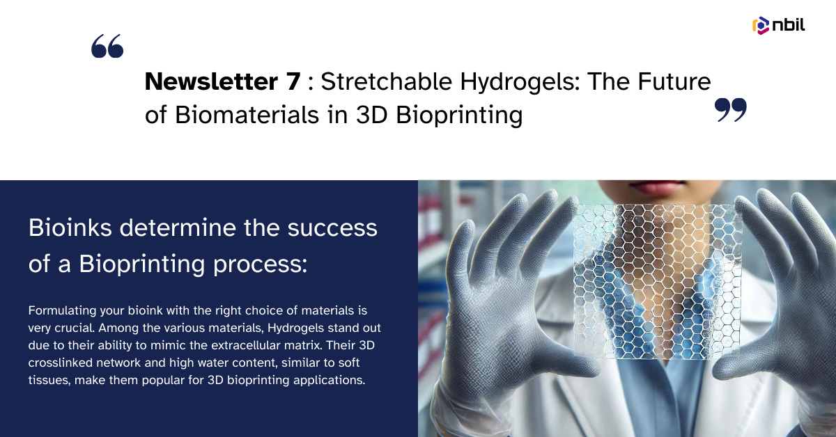
Bioinks determine the success of a Bioprinting process. Hence formulating your bioink with the right choice of materials is very crucial. Among the various materials, Hydrogels stand out due to their ability to mimic the extracellular matrix. Their 3D crosslinked network and high water content, similar to soft tissues, make them popular for 3D bioprinting applications.
Properties and Functionalities:
Hydrogels effectively replicate the extracellular matrix, the natural environment of cells, thus providing a hydrated and structurally supportive environment that can be homogeneously populated with cells.
Their hydrophilicity ensures biocompatibility, making them suitable for tissue construct fabrication. Furthermore, hydrogels support cell growth and are highly customizable, thus allowing control over cell functions like adhesion, migration, proliferation, and differentiation. A variety of cell types can thrive when encapsulated within hydrogels. In addition, the mechanical properties of hydrogels can be tailored to match the specific requirements of different tissues, ranging from soft to stiff. Many hydrogels are biodegradable, ensuring temporary support and eventual integration with host tissues.
The physicochemical properties of hydrogels, pH, temperature, and viscosity significantly influence their stability, functionality, and biocompatibility in 3D bioprinting. Most hydrogels are stored and processed at physiological pH (~7.4) to maintain structural integrity and minimize toxicity.
Temperature and viscosity have an inverse relationship: higher temperatures reduce viscosity. This can improve printability and reduce shear stress on embedded cells. However, the optimal printing temperature varies depending on the specific hydrogel material.
Crosslinking: Crosslinking, a post-printing process that enhances the mechanical properties of hydrogels, is typically performed at a physiological temperature (37°C) for natural polymer hydrogels. However, some hydrogels, such as alginate and gelatin, can undergo crosslinking at room temperature.
Applications in 3D Bioprinting:
Recent advancements in 3D bioprinting have made hydrogel-based systems ideal carriers for cells in tissue engineering and molecules or drugs in stem cell and cancer research.
Organ Fabrication: Hydrogels can be used to create functional organs, such as liver, kidney, and heart, for transplantation.
Tissue Engineering: Hydrogels can create tissue-engineered constructs for various applications, including wound healing, tissue development, and regenerative medicine.
Drug Delivery Systems: Hydrogels can be designed as drug delivery systems, releasing therapeutic agents in a controlled fashion.
Personalized Medicine: They can be customized to meet the specific needs of individual patients, thus enabling personalized treatments.
Despite advancements in hydrogel fabrication, traditional 3D-printed hydrogels may fracture when stretched or cracked under pressure. Additionally, some hydrogels are too rigid to conform to the shape of deformable tissues.
Interestingly, Researchers from the University of Colorado Boulder, in partnership with the University of Pennsylvania and the National Institutes of Standards and Technology (NIST), have developed a novel technique for fabricating 3D-printed hydrogels with enhanced mechanical properties and potential adhesive capabilities.
This innovative approach involves incorporating intertwined chains of molecules, resulting in hydrogels that exhibit superior strength, elasticity, and adhesion to wet tissues. The method, known as CLEAR, utilizes spatial light illumination (photopolymerization) to define an object's shape while simultaneously employing a complementary redox reaction (dark polymerization) to gradually build a dense network of entangled polymer chains.
CLEAR represents a groundbreaking advancement in biomaterial fabrication. It is the first method to successfully integrate light and dark polymerization using the Digital Light Processing Method (DLP). Furthermore, with no special equipment needed, CLEAR relies on conventional fabrication methods, with some tweaks in processing.
From a literature survey, the group found that these entangled polymeric network hydrogels are often made with slow reactions that are not compatible with DLP, as each layer reacts through shorter durations of light. This limitation was overcome using DLP with light and slow redox dark polymerization.
Experimental results demonstrated that hydrogels fabricated using the CLEAR technique were significantly more robust, exhibiting a four to seven times greater toughness than those produced through traditional DLP methods. Additionally, CLEAR-produced hydrogels demonstrated the remarkable ability to mold and adhere to complex organs, as illustrated by its successful integration with a porcine heart.
The researchers emphasize the potential of CLEAR-printed hydrogels as versatile tissue adhesives. By fabricating these adhesives into diverse structures, such as porous lattices or spatially defined patterns, CLEAR offers promising solutions for biomedical applications. Moreover, CLEAR's applications extend beyond hydrogels, suggesting its potential for broader integration into manufacturing processes.
Hence, CLEAR has the potential to advance biomedical research and reduce the environmental footprint of manufacturing and research processes by eliminating the need for additional energy/light sources and focusing on sustainability.
Future Directions:
The researchers focus on identifying specific applications where CLEAR can make the most significant impact and conducting further studies to understand its long-term effects on tissues.
Stay tuned for our next edition, where we will provide exclusive updates on bioprinting.
Reference-
https://physicsworld.com/a/3d-printing-creates-strong-stretchy-hydrogels-that-stick-to-tissue/
https://www.ncbi.nlm.nih.gov/pmc/articles/PMC10339415/
Curated Biotechnology Jobs:
