

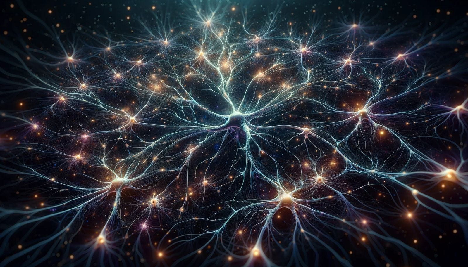
Unravel the Complexities with the First 3D Printed Brain Tissue
Welcome to our second edition of the newsletter, In this edition, we'll be unveiling the groundbreaking work on 3D Printed Brain Tissue by Pioneers at the University of Wisconsin, Madison, USA.

Unlock the Intricacies of the Brain through Comprehensive Insights and Perspectives.
Understanding the functioning of human neural networks encounters challenges due to the scarcity of dependable human neural tissues that can facilitate dynamic evaluations of neural circuits. 3D bioprinting offers a more precise and controlled method for creating human neural tissues, as it involves meticulous placement of living cells and hydrogels to construct biologically intricate cytoarchitecture using human pluripotent stem cells, induced pluripotent stem cells (iPSCs), and human embryonic stem cells (hESCs). Various culture systems, including hydrogel cultures, scaffold-based cultures, acoustic levitational assembly, self-organized brain spheroids/organoids, and brain-on-a-chip models, have been employed to engineer these complex neural structures.
Previous studies employed 3D printers to create brain tissues, facilitating researchers' examination of cell maturation, connection formation, and even the integration of printed tissue into mouse brains. However, these constructs possessed limited functionality. Research endeavors that produced more functional printed tissues utilized rat cells instead of human cells.
In other cases, the scaffold fabricated through 3D printing followed the seeding of cells onto it. Unfortunately, neural cells within these structures often formed uneven distributions, leading to large, thick cell clusters. Challenges arise from the scaffold's non-biodegradable nature, which hinders neural cell migration and impairs the formation of neural networks between tissue layers. The 3D bioprinted neural tissues were fabricated using soft gels such as gellan gum, alginate, or gelatin mixed with fibrin that exhibited layered structures with a degree of neuronal maturation. However, when printed in a layer-by-layer manner vertically, the functional neuron-neuron or neuron-glial connections within or between tissue layers remained unproven.
Interestingly, researchers from the University of Wisconsin-Madison, USA, introduced a technology platform that enables the assembly of neuronal and glial subtypes into precisely structured 3D neural tissues. This innovative approach facilitates the formation of functional connections between neurons and glial cells within and between tissue layers, employing extrusion bioprinting. Unlike traditional methods that stack layers vertically, this technique prints layers or bands horizontally adjacent to each other. These uniquely designed 3D neural tissues can be sustained using conventional culture systems, offering ease in live-cell imaging and electrophysiological recording. This groundbreaking platform opens new avenues for studying human neural networks under both physiological and pathological conditions.
Crucial Elements for Successful Bioprinting: The Bioink's Role in Cell Survival and Neural Maturity
The core of successful bioprinting procedures is the bioink. It ensures cell preservation, fosters proper neural cell development, uniform cell distribution, adjustable gelation timing, and provides strong structural stability. Fibrin hydrogel, primarily composed of fibrinogen and thrombin, has proven to be biocompatible with neural cells. Consequently, Zhang's team opted for fibrin gel as the primary bioink for their printing method, ensuring an ideal environment for neural tissue growth. While fibrin gel is well-suited for neural cell compatibility, its high viscosity presents challenges in bioprinting procedures. To tackle this issue, a printable bioink was formulated by merging fibrin gel with hyaluronic acid, thereby enhancing its compatibility for neural tissue printing. Additionally, this combination demonstrated support for cortical neuron maturation and synaptogenesis.
The Art of 3D Bioprinting lies in its Design Considerations
The researchers have employed extrusion-based bioprinting to deposit gels layer by layer, allowing for the replication of intricate structures, such as the intricate laminations found within the human cortex.
Thicker neural tissues can hinder oxygen and nutrient transfer, negatively impacting printed neural cell growth and function without a vascular system. As such, optimal printed brain tissue thickness should ideally fall within the 100-200 µm range. To address ease of assessment, broad applicability, and compatibility with standard laboratories, Zhang's team opted for a layer thickness of 50 µm.
The study developed a novel tissue layout in 3D bioprinting: Horizontal Band Printing for Enhanced Analysis, instead of the conventional vertical stacking method. They meticulously printed separate horizontal lines of human neural and glial progenitor cells, which have the potential to develop into diverse brain cell types. This design enables the creation of thin, multi-layered, functional neural tissues with defined cellular compositions and desired dimensions. These tissues can be effortlessly maintained and assessed in routine laboratory settings.
The resulting 3D structures accurately mirrored the development of brains, as cells established connections within their respective bands and extended links towards other bands. This unique setup enabled researchers to study the maturation process and interlinking of progenitor cells.
This tailored brain tissue design offers a well-defined platform for investigating human neural networks under both physiological and pathological conditions.
To further augment the capabilities of their 3D bioprinted neural tissues, the research team meticulously designed and synthesized diverse constructs by integrating various cell types with specific ratios. One such configuration comprised inhibitory and excitatory neurons, which communicate through distinct types of signaling molecules called neurotransmitters. Furthermore, essential supporting astrocytes were incorporated into these constructs. In these advanced neural constructs, neurons often generated electrical signals, and astrocytes efficiently absorbed the neurotransmitter glutamate, demonstrating the formation of functional connections akin to those found in the human brain. When the researchers combined cell types representative of the brain's outer cortex and deeper striatum, they observed that cortical cells extended projections towards striatal cells, but not vice versa. This observation mirrored the organization found in the human brain, confirming the constructs' potential to replicate brain organization.
Challenges and limitations
The gel-based bioink's softness restricts vertical multi-layer printing capabilities, leading to a thickness constraint of approximately 50 mm for printed tissue. Moreover, the technology does not yet support the orientation of mature neurons, as many exhibit a pyramidal shape, resulting in a lack of inherent structural organization compared to brain organoids.
Despite these constraints, the printed tissue retains some intrinsic properties, such as anatomical and functional neuronal connections, as demonstrated by unidirectional axonal projection from cortical to striatal tissue. The printed brain tissue exhibits rapid, synchronized cell maturation, complementing existing organoids and providing a controlled environment for studying human neural networks under both physiological and pathological conditions.
In Conclusion…
This work holds immense significance for neuroscientists investigating the brain and working towards the advancement of treatments for a multitude of neurological and neurodevelopmental disorders, such as Alzheimer's and Parkinson's disease, conducting drug tests, and even exploring brain growth. It can be utilized to study a wide range of key aspects, such as molecular mechanisms in brain development, human development, developmental disabilities, neurodegenerative disorders, and more.
As bioprinting technology continues to evolve, it is expected that more complex and accurate neural tissue models will be produced, paving the way for enhanced understanding of human neural circuits and their applications in neuroscience research.
Stay tuned as we endeavor to provide you with our next edition with exclusive updates in the field of Bioprinting.